https://doi.org/10.1038/nature18933
- paper gelesen?
- Infos rausgeschrieben?
Glasser et al. (2016) - Nature
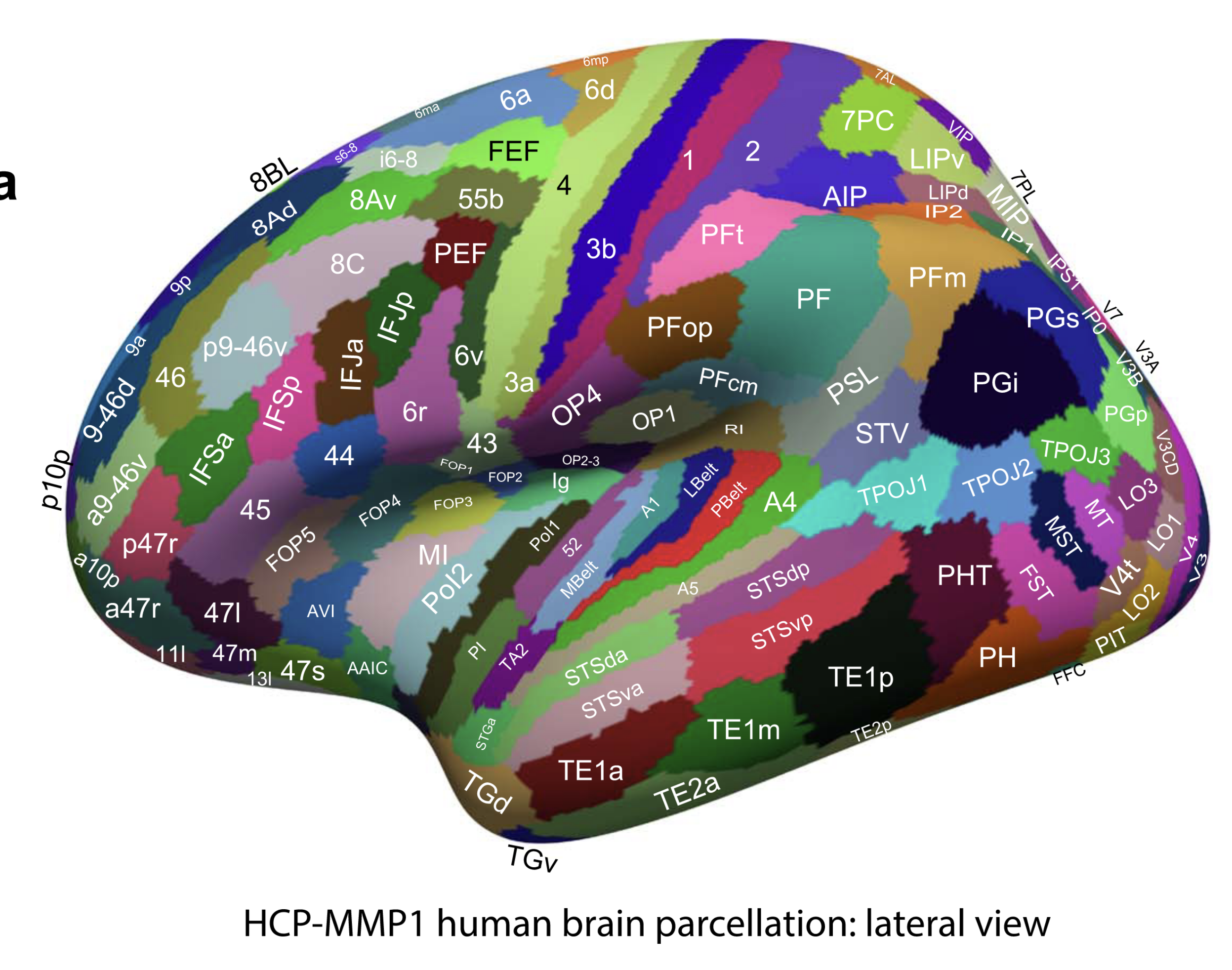
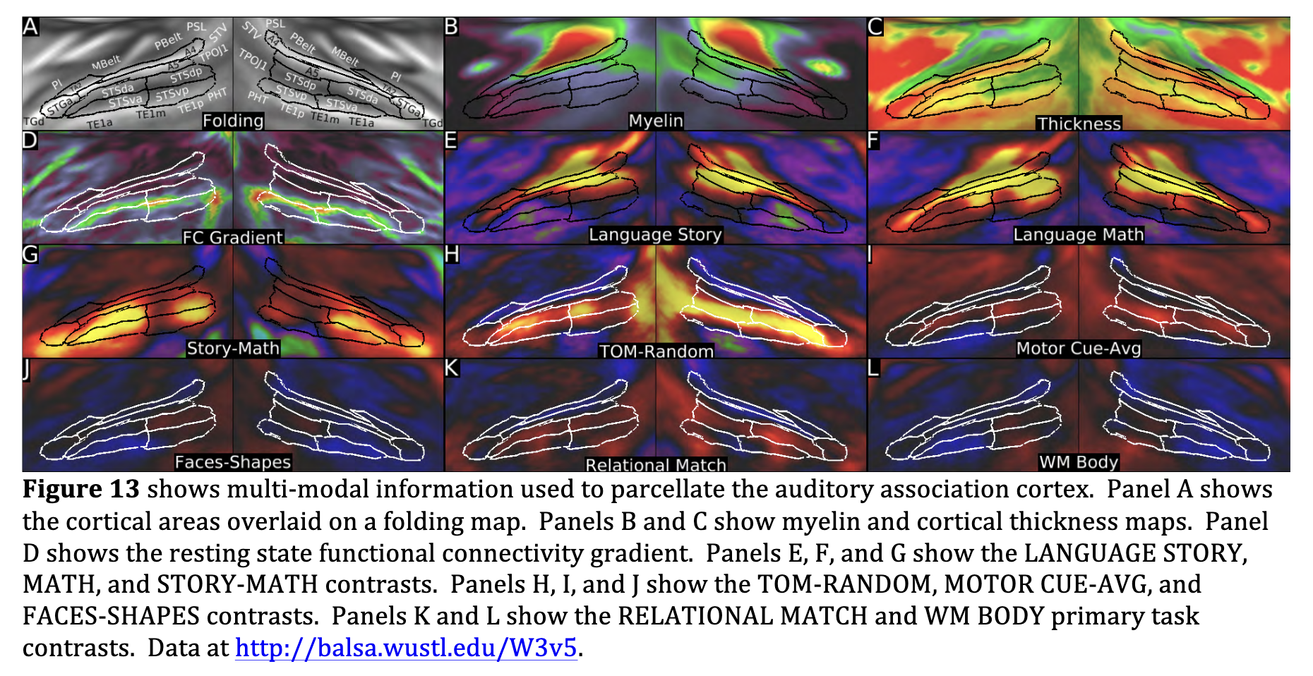
- The early auditory areas include A1, LBelt (Lateral Belt), MBelt (Medial Belt), PBelt (Para-Belt), and the retro-insular cortex (RI). These areas are surrounded by areas, OP2-3, OP1, PFcm, PSL, A4, Ig, and TA2.
- A1 is very heavily myelinated, even relative to its heavily myelinated surrounding neighbors
- interesting colour asymmetries occur in a few areas, especially language-related areas 55b, PSL, SFL, and 44 and their right hemisphere homologues, which also have asymmetric task-fMRI functional profiles
⇒ jetzt verstehe ich auch, warum die Belt regions so langgezogen sind. Sie umgeben einfach den A1 - das macht sie so langgezogen uns der ganze bereich hat eben sehr ähnliche connectivity.
- Relative to its antero-medial neighbor area 52, the MBelt complex has more myelin (Panel B), is thicker (Panel D), and is activated vs deactivated in the LANGUAGE MATH and STORY contrasts (Panel J). Relative to its antero-lateral neighbor area TA2, MBelt has more myelin (Panel B), is thinner (Panel D), and is more activated in the LANGUAGE MATH and STORY contrasts (Panel J). Relative to its lateral neighbor PBelt, the MBelt complex has more myelin (Panel C) and is less activated in the language MATH and STORY contrasts (Panel J) and more activated in the EMOTION FACES-SHAPES contrast.
- We identified auditory association cortex as a region mainly on the superior temporal gyrus and within the superior temporal sulcus that is activated in the LANGUAGE STORY, MATH, and STORY-MATH contrasts.
- It is strongly functionally connected with the inferior frontal gyrus, including areas 44, 45, and 47l.
- This auditory region likely becomes progressively less purely auditory and more multi-modal as one progresses inferiorly, anteriorly, and posteriorly (away from early auditory cortex, e.g. Main Text Figure 3).
- A4, A5, STSdp, STSda, STSvp, STSva, STGa, and TA2
- we have introduced largely novel terminology here, except that TA2 is based on the Von Economo and Koskinas parcellation (Triarhou, 2007a, b; von Economo and Koskinas, 1925). These areas are surrounded by PBelt, MBelt, PI, TGd, TE1a, TE1m, TE1p, PHT, TPOJ1, STV, and PSL.
more about A4:
A4’s supero-medial border with PBelt was covered in Section #10 Early Auditory Cortex. Relative to its inferior neighbor A5, area A4 differs in functional connectivity, and this gradient was primarily used to define the boundary (Panel D). A4 also has more myelin than A5 assessed statistically (Panel B), though the myelin gradient peak does not align with the functional connectivity gradient peak. Relative to its antero-medial neighbor TA2, area A4 has more myelin (Panel B), differs in functional connectivity (Panel D), is more activated in the LANGUAGE MATH and STORY contrasts (Panels E and F), and is less activated in the CUE-AVG contrast (Panel I).
more about A5
- A5 differs in functional connectivity (Panel D), and is more activated in the LANGAUGE STORY-MATH (Panel G) and TOM-RANDOM (Panel H) contrasts. Relative to its inferior neighbor STSdp, area A5 differs in many primary and non-primary task contrasts including more activation in the LANGAUGE MATH (Panel E) and STORY (Panel F) contrasts and less activation in the working memory (e.g Panel L) and RELATIONAL (e.g. Panel J) primary contrasts, and the FACE-AVG, TOM-RANDOM (Panel H), and FACES-SHAPES (Panel K) contrasts. Relative to its inferior neighbor STSda, area A5 again shows differences in a variety of task contrasts including markedly more activation in the LANGAUGE MATH contrast (Panel E), more activation in the TOOL-AVG contrast, markedly less activation in the TOM-RANDOM contrast (Panel H), and less activation in the CUE-AVG (Panel I) and FACES-SHAPES (Panel K) contrasts. Relative to its anterior neighbor STGa, area A5 has more myelin (Panel B) and differs in functional connectivity (Panel D).
more about STSdp:
- Relative to area STSvp on the inferior bank of the STS, area STSdp on the superior banks is has more myelin (Panel B), differs markedly in functional connectivity (Panel D), is more activated in the LANGUAGE MATH (Panel E), TOM-RANDOM (Panel H, especially in the right hemisphere), and MOTOR CUE-AVG (Panel I) contrasts. Relative to its anterior neighbor STSda in the superior bank of the STS, area STSdp has more myelin (Panel B), and differs markedly in its functional activation profile, being more activated in the CUE-AVG contrast (Panel I) and the RELATIONAL MATCH (Panel J), working memory (e.g. Panel L), SOCIAL TOM and other primary contrasts, and less active in the STORY-MATH contrast (Panel G).
chatbot content following:
careful with using this information
from SUPPL about STV:
STV
- Distinctive Features: Both STV and PSL possess unique features beyond typical multi-modal gradients and show prominent asymmetries between hemispheres.
- STV (Superior Temporal Visual area):
- Defined using gradients from functional connectivity maps seeded from the PCV (Posterior Cingulate Visual area).
- Shows strong functional connectivity with the PCV.
- PSL (Peri-Sylvian Language area):
- Defined using gradients from functional connectivity maps seeded from the unilateral Area 55b.
- Contains an anterior-to-posterior topographic organization of connectivity (similar to areas 55b, SFL, and 44)
- Border with Area A4:
- Moderately long in the Left Hemisphere.
- Almost nonexistent in the Right Hemisphere.
- Relative to Area A4 (Left Hemisphere):
- STV is more connected with PCV than A4 is.
- Deactivated vs. activated in: TOOL-AVG, LANGUAGE MATH, and STORY contrasts.
- More activated in: MOTOR CUE-AVG contrast.
- Activated vs. deactivated in: SOCIAL primary contrasts and TOM-RANDOM contrast.
- Relative to TPOJ1 (Inferior Neighbor):
- STV has less myelin.
- STV has stronger functional connectivity with PCV.
- STV is deactivated vs. activated in LANGUAGE MATH and STORY contrasts.
- Left Hemisphere: STV is less activated than TPOJ1 in GAMBLING primary contrasts and MOTOR AVG.
- Right Hemisphere: STV is less activated than TPOJ1 in TOM-RANDOM contrast.
- Relative to PGi (Posterior Neighbor):
- Differs strongly in functional connectivity.
- Less activated in LANGUAGE STORY and STORY-MATH contrasts.
- More activated in SOCIAL RANDOM contrast.
- Relative to PFm (Posterior Neighbor):
- Differs strongly in functional connectivity.
- More activated in SOCIAL primary contrasts.
- Relative to PSL (Superior Neighbor):
- STV has stronger connectivity with PCV, whereas PSL has stronger connectivity with 55b
- STV is deactivated (vs. PSL activated) in: Working memory primary contrasts, MOTOR AVG (Panel L), LANGUAGE MATH, STORY (LH only), and RELATIONAL primary contrasts.
from SUPPL about PSL:
PSL
- Relative to Area A4 (Antero-inferior Neighbor):
- Stronger functional connectivity with Area 55b.
- Activated vs. deactivated in primary contrasts.
- Deactivated vs. activated in TOOL-AVG contrast.
- More activated in MOTOR AVG.
- Relative to PFm (Supero-posterior Neighbor):
- Stronger functional connectivity with Area 55b.
- More activated in RELATIONAL primary contrasts (bilateral).
- Left Hemisphere: More activated in LANGUAGE MATH and STORY contrasts.
- Right Hemisphere: More activated in MOTOR CUE and TOM-RANDOM contrasts.
- Relative to PF (Supero-anterior Neighbor):
- Stronger functional connectivity with Area 55b.
- Left Hemisphere: Less activated in MOTOR CUE; more activated in LANGUAGE MATH and STORY.
- Right Hemisphere: Less deactivated in LANGUAGE contrasts; more activated in TOM-RANDOM (but not on the Left).
4. Lateralization and Topography of PSL
-
Lateralization:
-
PSL is one of the few “strikingly functionally lateralized” areas in the cortex.
-
Cortical Thickness: PSL is much thicker on the Right than on the Left.
-
Activation: Left PSL is more activated in RELATIONAL-MATCH contrasts than Right PSL.
-
Geography: Located near the supero-posterior tip of the Sylvian fissure, extending onto the Superior Temporal Gyrus (especially on the Right).
-
-
Topographic Organization (Language Network):
-
PSL, 55b, SFL, and 44 show a topographic organization of connectivity along the anterior-posterior axis.
-
This pattern is part of a left-lateralized language Resting State Network (RSN).
-
Left Hemisphere Gradient Mapping: Progression from Posterior to Anterior in PSL corresponds to:
-
Anterior to Posterior in Area 55b.
-
Inferior to Superior in Area 44.
-
Anterior to Posterior in SFL (more prominent on the Left).
-
-
We identified the temporo-parieto-occipital junction as a strip of cortex bounded by auditory, lateral temporal, inferior parietal and occipital (visual MT+ complex) regions. This region contains five multimodal areas, TPOJ1, TPOJ2, TPOJ3, STV, and PSL, that are surrounded by areas PGp, PGi, PFm, PF, PFcm, RI, A4, A5, STSdp, STSvp, PHT, FST, MST, MT, and LO3. Areas TPOJ1-3 are moderately myelinated and show strong functional connectivity among themselves and with STV. They form a bridge between higher auditory and higher visual areas, as they are correlated with both. Figure 17 shows the multi-modal information used to parcellate these areas, along with the areas on a folding map (Panel A). All five areas have novel names, as these areas to do not clearly correspond with previous parcellations of this region.
OP4’s boundary with area 1 was covered previously in Section #6 Somatosensory and Motor Cortex. Relative to its supero-posterior neighbor PFop, area OP4 differs in functional connectivity (Panel D) and is modestly activated vs strongly deactivated in the LANGUAGE STORY contrast (Panel F).
Paper
Supplementary work
⇒ Was bedeuten die Vergleiche aus Glasser SUPPL
see also
Tags: neuroscience science source
Superlink: 050 🧠Neuroscience
Created: 2025-11-12 22:14